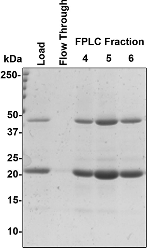
FIGURE 3.

Purified SN-Hendra F protein. Samples from load, flow-through, and the three FPLC fractions containing the majority of the Hendra SN-TM protein were separated on a 15% SDS-PAGE gel and visualized by Coomassie Blue Staining. Bands corresponding to the monomeric and an oligomeric size are apparent.
