FIGURE 2.
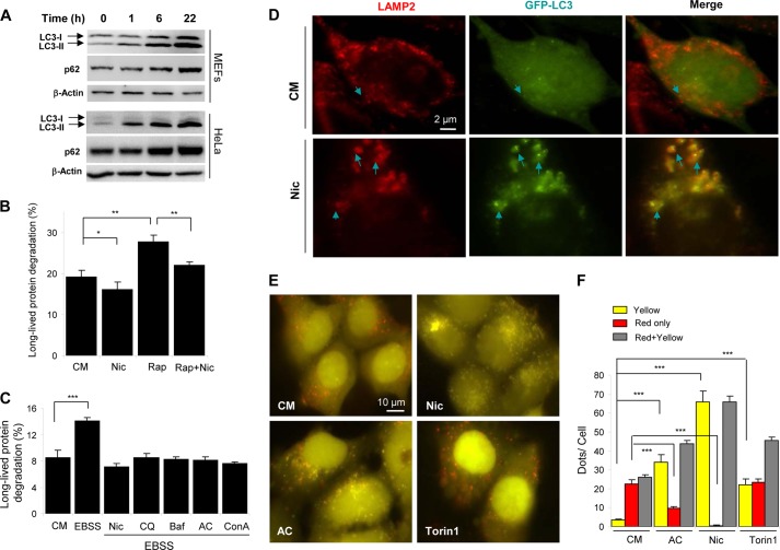
Niclosamide inhibits autophagic degradation. A, MEFs or HeLa cells were treated with niclosamide (Nic; 10 μm) for 0–22 h as indicated, followed by immunoblot analysis for p62 and LC3. B and C, MEFs were incubated in complete medium (CM), or in Earle's balanced salt solution (EBSS) with or without niclosamide (10 μm), CQ (40 μm), Baf (0.5 μm), AC (40 mm) or ConA (2.5 μm) for 6 h (C), or incubated in complete medium (CM) with or without niclosamide (10 μm) and rapamycin (Rap; 1 μm) for 16 h (B). The long-lived protein degradation was measured. D, GFP-LC3-expressing MEFs were treated as indicated (Nic, 10 μm) for 6 h, followed by immunostaining with anti-LAMP2. Arrows indicated colocalized GFP-LC3 (green) and LAMP2 (red) puncta. E and F, HEK-293A cells expressing GFP-RFP-LC3 were treated as indicated (Nic, 10 μm; AC, 40 mm; or Torin1, 250 nm) for 6 h (E). Puncta showing both green and red fluorescence (indicated as yellow) or showing only the red fluorescence (indicated as red) were quantified (F). Values represent means ± S.D. from three independent experiments. ***, p < 0.001; **, p < 0.01; *, p < 0.05.

