FIGURE 3.
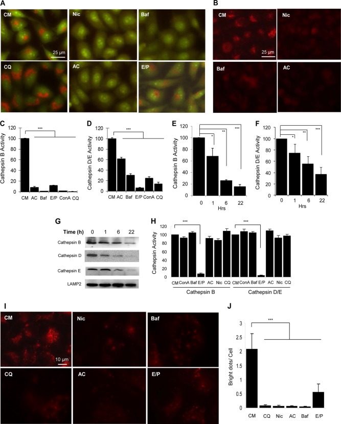
Niclosamide inhibits lysosomal degradation capacity. A and B, HeLa cells were treated with niclosamide (Nic; 10 μm), CQ (40 μm), Baf (0.5 μm), AC (40 mm), or E64D (25 μm) plus pepstatin A (50 μm) (E/P) for 6 h, followed by staining with AO (1 μg/ml, A) or LTR (50 ng/ml, B) for 30 min. C–G, HeLa cells were treated with various chemicals (40 mm AC; 0.5 μm Baf; 25 μm/50 μm E64D/pepstatin A; 2.5 μm ConA; 40 μm CQ) as indicated (C and D) for 6 h, or with niclosamide (10 μm) for 0 to 20 h (E–G). The lysosome-enriched cytosolic fraction was analyzed for cathepsin B (C and E) and cathepsin D/E (D and F) activities and for immunoblot analysis (G). Cathepsin activities were standardized to that of the untreated sample, which was set at the 100% level. H, whole cell lysates of untreated HeLa cells (4 μg) were mixed with the various chemicals at the concentrations indicated above for 1 h. The activities of cathepsin B and cathepsin D/E were measured. I and J, HeLa cells were preincubated with DQ-BSA(10 μg/ml) for 1 h and then treated as in A (I). The degraded products presented as red puncta, which were quantified (J). For C–F, H, and J, values represent means ± S.D. from three independent experiments. ***, p < 0.001; **, p < 0.01; *, p < 0.05. CM, complete medium.

