
FIGURE 3.
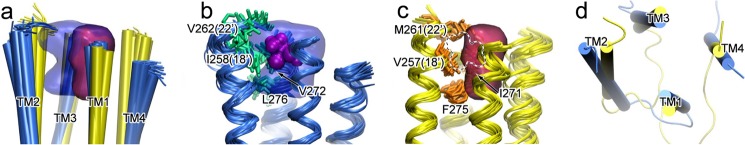
The intrasubunit cavity at the EC end of the TM domain in β2, but not in α7, can accommodate isoflurane binding. a, alignment of 20 NMR structures with the lowest target function for β2 (blue) and α7 (yellow), and the cavities of β2 (blue) and α7 (red), outlined by grid points present in at least five of the 20 structures. b and c, residues highlighted with the side chain bundles (shown in stick representation) in β2 (b) and α7 (c) have primary responsibility for the different cavity volumes. Note that in β2, the cavity can accommodate isoflurane (purple surface), but the cavity in α7 (dotted outline) cannot do the same. d, the top view of the lowest target function structures of β2 (blue) and α7 (yellow) shows different orientations of TM helices.