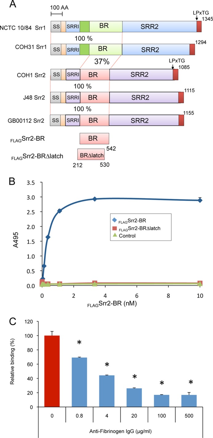
FIGURE 2.

Interaction of the BR of Srr2 with fibrinogen. A, schematic diagram of the serine-rich repeat proteins Srr1 and Srr2. Level of identity (%) between regions is indicated. SS, signal sequence; Srr1-BR and Srr2-BR, binding domains; SRR1 and SRR2, serine-rich regions; LPxTG, cell wall anchoring motif; AA, amino acids. B, binding of FLAG-Srr2-BR and FLAG-Srr2-BRΔlatch proteins to immobilized fibrinogen. Indicated concentrations of FLAG-Srr2-BR and FLAG-Srr2-BRΔlatch were added to wells coated with fibrinogen or casein blocking reagent. C, inhibition of FLAG-Srr2-BR binding to immobilized fibrinogen by anti-fibrinogen IgG. Values represent percent of FLAG-Srr2-BR binding to the wells treated with fibrinogen. Bars indicate the means ± S.D. *, p < 0.01.
