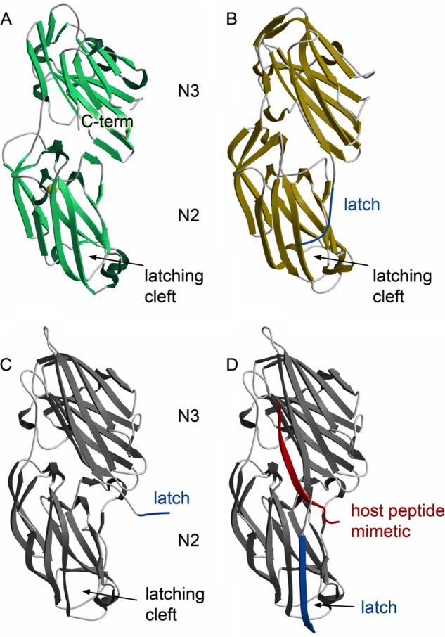
FIGURE 3.

Structures of Srr1-BR and Srr2-BR. A, structure of Srr1-BR with secondary structural elements colored green and turns colored in gray. The latch region is disordered in the structure. B, structure of Srr2-BR with secondary structural elements colored yellow and turns colored gray. The latch region is highlighted in cerulean blue. C, structure of ClfB without peptide ligand mimetic shows a latching region that is open and an unoccupied latching cleft ((Protein Data Bank entry 4F24 (19)). The ordered region of the latch is highlighted in cerulean blue. D, structure of ClfB with peptide ligand mimetic identifies peptide binding to the cleft between the N2 and N3 domains and shows the latch in the locked position. (Protein Data Bank entry 4F27 (19)). The peptide is shown in red, and the latch is shown in cerulean blue.
