FIGURE 3.
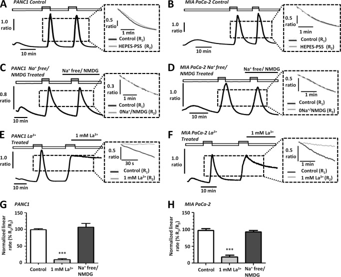
PMCA is the main mechanism of [Ca2+]i efflux in human PDAC cell lines. A–F, representative traces show the in situ [Ca2+]i clearance assay (PMCA activity) for control PANC1 (A) and MIA PaCa-2 (B) cells, Na+-free/NMDG-treated PANC1 (C) and MIA PaCa-2 (D) cells, and La3+ treated PANC1 (E) and MIA PaCa-2 cells (F). Cells were treated with CPA (30 μm) in zero external Ca2+ with 1 mm EGTA (white box) or 20 mm Ca2+ (gray box) to induce store-operated Ca2+ influx. Subsequent removal of external Ca2+ resulted in [Ca2+]i clearance. This influx-clearance phase was repeated under conditions where extracellular Na+ was replaced with equimolar Na+-free/NMDG or in the presence of 1 mm La3+. La3+ was prepared in HEPES-PSS devoid of EGTA to prevent chelating of the La3+. The inset of each trace shows expanded time courses comparing the second (gray trace) with the first clearance phase (black trace) in the presence of each treatment. The linear clearance rate over 60 s (in the presence of each treatment) was normalized to the initial clearance rate in each cell (% relative clearance). Normalized linear rate (± S.E.) is presented for PANC1 (G) and MIA PaCa-2 (H) cells. ***, p < 0.001 (Mann-Whitney U test) compared with time-matched control experiments (white bar).

