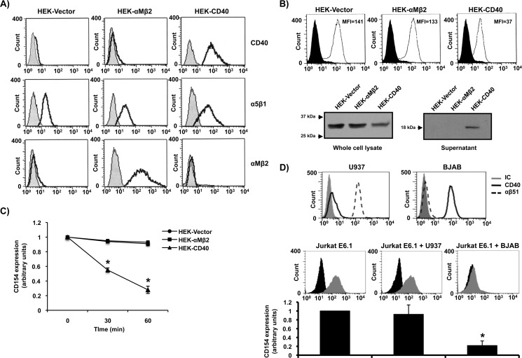
FIGURE 1.
Membrane-bound CD154 is cleaved upon its interaction with CD40. A, expression profiles of CD40, α5β1, and αMβ2 on HEK293-Vector, HEK293-αMβ2, and HEK293-CD40 cells are shown. Cells (5 × 10⁶/ml) were stained with the appropriate antibody (CD40, clone G28.5; α5β1, clone JBS5; and αMβ2, clone ICRF44) or its isotype control (IC) for 1 h at 4 °C, washed, and incubated with an anti-mouse IgG Alexa Fluor 488-coupled secondary antibody for 30 min at 4 °C. Sample were then washed and analyzed by FACS analysis. Histogram plots are representative of four independent experiments. B, Jurkat E6.1-CD154 cells (10 × 106/ml) were co-cultured with HEK-Vector (10 × 106/ml), HEK-αMβ2 (10 × 106/ml), or HEK-CD40 (10 × 106ml) cells for 1 h at 37 °C. Total cell lysates and supernatants were then analyzed by Western blotting for CD154 expression (clone C4.14). Full-length membrane-bound CD154 (≈33 kDa) is shown in whole cell lysates, whereas sCD154 (≈18 kDa) is found within the supernatants from co-cultures with HEK-CD40 cells only. Upper histogram plots show representative CD154 shedding at 60 min of co-culturing experiments as described in C. C, Jurkat E6.1 cells stably transfected with CD154 (5 × 10⁶/ml) were co-cultured with HEK293 cells (5 × 10⁶/ml) stably transfected with vector alone (HEK-Vector), with αMβ2 (HEK-αMβ2), or with CD40 (HEK-CD40) for the indicated time at 37 °C. Cells were then stained with an anti-CD154 antibody (clone C4.14) or its isotype control for 1 h at 4 °C, washed, and incubated with an anti-mouse IgG Alexa Fluor 488-coupled secondary antibody. CD154 shedding was then measured by FACS analysis as arbitrary units of residual membrane expression over time. D, upper representative histogram plots show the expression of α5β1 (clone JBS5) and CD40 (clone G28.5) on both U937 and BJAB cells. Cells (5 × 10⁶/ml) were stained as described in A and analyzed by FACS analysis. Lower plots and histogram bars of mean of data (n = 3; *p < 0.05 versus Jurkat E6.1 alone) show the impact of U937 and BJAB cells on CD154 cleavage from Jurkat E6.1-CD154 cells. Jurkat E6.1 cells stably transfected with CD154 (5 × 10⁶/ml) were co-cultured with U937 (5 × 10⁶/ml) or BJAB (5 × 10⁶/ml) cells or left alone for 1 h at 37 °C. Cells were then double stained with an anti-CD3-FITC antibody (clone UCHT1) and an anti-CD154-biotin-conjugated antibody (clone C4.14) or their respective isotype controls for 1 h at 4 °C. Samples were then washed and incubated with streptavidin-phycoerythrin for 30 min at 4 °C for CD154 fluorescence. Cells were washed, and double fluorescence was detected by FACS analysis using the appropriate compensation parameters. Histogram plots are representative of CD154 fluorescence within the CD3-positive population (Jurkat E6.1 T-cells).

