FIGURE 4.
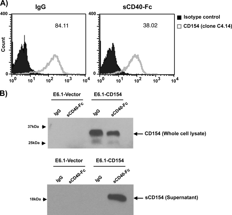
Soluble CD40 induces CD154 shedding. A, Jurkat E6.1-CD154-transfected cells (5 × 10⁶/ml) were stimulated with soluble CD40-Fc (1.25 μg/ml) or negative control IgG (1.25 μg/ml) for 1 h at 37 °C. Cells were then washed and stained with an anti-CD154 antibody (clone C4.14) for 1 h at 4 °C. Samples were thereafter incubated with an anti-mouse IgG Alexa Fluor 488-coupled secondary antibody for 30 min at 4 °C, washed, and analyzed by FACS for membrane-bound CD154 expression. Histogram plots are representative of five independent experiments (values within plots represent the mean fluorescence intensity for each sample). B, Jurkat E6.1-Vector-transfected (10 × 106/ml) and Jurkat E6.1-CD154-transfected (10 × 106/ml) cells were stimulated with sCD40-Fc (1.25 μg/ml) or negative control IgG (1.25 μg/ml) for 1 h at 37 °C. Total cell lysates and supernatants were then analyzed by Western blotting for CD154 expression (clone C4.14). Full-length membrane-bound CD154 (≈33 kDa) is shown in whole cell lysates, whereas sCD154 (≈18 kDa) is found within the supernatants of E6.1-CD154 cells stimulated with sCD40-Fc only. Blots are representative of four independent experiments.

