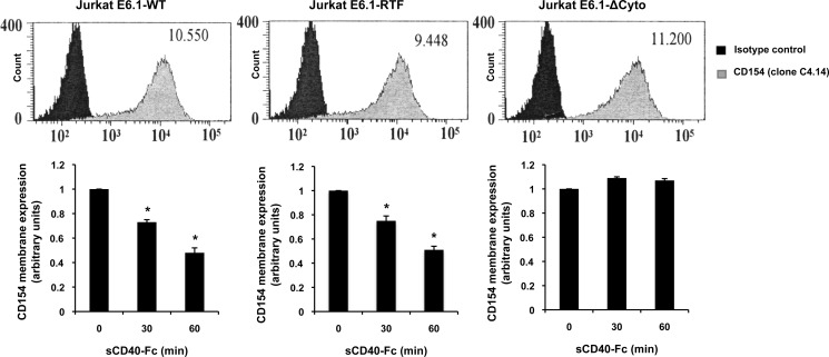
FIGURE 5.
CD154 shedding is independent of lipid raft integrity. Jurkat E6.1-CD154 wild-type transfected (Jurkat E6.1-WT), Jurkat E6.1-CD154 transferrin receptor I mutant (Jurkat E6.1-RTF), and Jurkat E6.1-CD154 cytoplasmic domain deletion mutant (Jurkat E6.1-ΔCyto) cells were assessed for CD154 cell surface expression by FACS analysis (upper histogram panels). Cells (5 × 10⁶/ml) were stained with an anti-CD154 antibody (clone C4.14) or its isotype control for 1 h at 4 °C, washed, and incubated with a secondary anti-mouse IgG Alexa Fluor 488-coupled antibody for an additional 30 min at 4 °C. All histogram plots are representative of four independent experiments (values within plots represent the mean fluorescence intensity). Lower histogram bars represent the mean of data (n = 4; *p < 0.05; error bars, S.E.) of CD154 cell surface cleavage for all three populations. Cells (5 × 10⁶/ml) were stimulated with sCD40-Fc (1.25 μg/ml) for the indicated time, washed, and stained with an anti-CD154 antibody (clone C4.14) or its isotype control for 1 h at 4 °C. Samples were then washed and incubated with a secondary anti-mouse IgG Alexa Fluor 488-coupled antibody for an additional 30 min at 4 °C. Cleavage was assessed by FACS analysis, and results are expressed as arbitrary units of CD154 membrane expression in the absence (time 0) or presence of sCD40-Fc (30- and 60-min stimulation).

