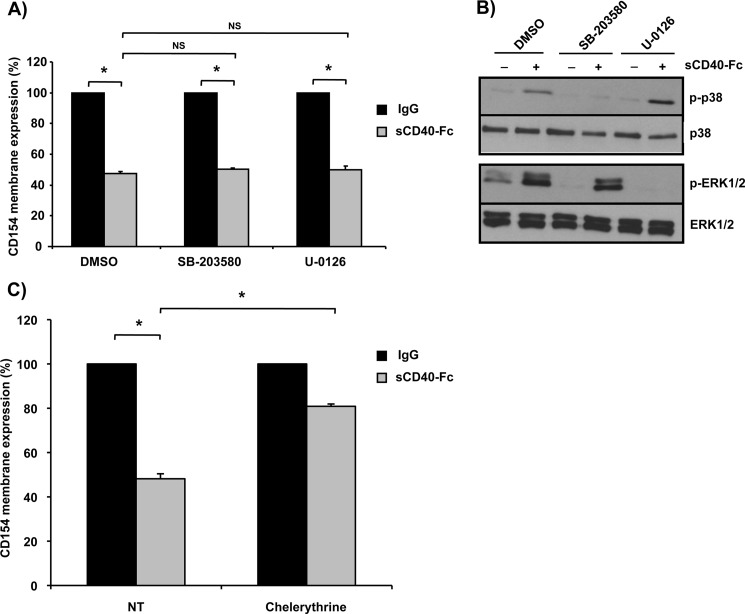
FIGURE 6.
CD154 shedding is modulated by PKC activation. A, Jurkat E6.1-CD154-transfected cells (5 × 10⁶/ml) were preincubated with the p38 SB203580 inhibitor (20 μm), the ERK1/2 U0126 inhibitor (20 μm), or control dimethyl sulfoxide (DMSO) for 15 min at 37 °C. Cells were then stimulated with sCD40-Fc (1.25 μg/ml) or negative control IgG (1.25 μg/ml) for an additional hour at 37 °C. CD154 shedding was assessed by residual cell surface expression by FACS analysis as outlined previously (n = 3; *p < 0.05 and NS = nonsignificant; error bars, S.E.). B, Jurkat E6.1-CD154 transfected cells (5 × 10⁶/ml) cells were preincubated with inhibitors as described in A and subsequently stimulated with sCD40-Fc (1.25 μg/ml) or not for 5 min at 37 °C. Samples were then lysed and phospho-p38 (p-p38), phospho-EKR1/2 (p-ERK1/2), as well as total p38 (T-p38) and total ERK1/2 (T-ERK1/2) were analyzed by Western blotting. Blots are representative of four independent experiments. C, Jurkat E6.1-CD154-transfected cells (5 × 10⁶/ml) were stimulated with sCD40-Fc (1.25 μg/ml) or negative control IgG (1.25 μg/ml) for 1 h at 37 °C in the absence (control dimethyl sulfoxide) or presence of the PKC inhibitor, chelerythrine (1 μm). CD154 shedding was thereafter assessed by FACS analysis as outlined previously (n = 4; *, p < 0.05).

