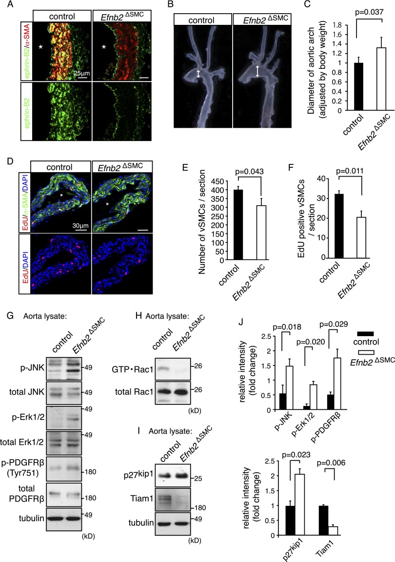
Figure 1.
Vessel wall defects in smooth muscle cell-specific ephrin-B2 mutants. (A) Confocal images showing ephrin-B2 (green) and α-smooth muscle actin (α-SMA; red) immunostaining on sections of adult (30 wk) control and Efnb2ΔSMC mutant aortae. (*) Vessel lumen. (B) Dilation affecting freshly isolated adult Efnb2ΔSMC aortic arches (right) compared with control littermates (left). Arrows indicate vessel diameter. (C) Quantitation of relative aortic arch diameter of adult mice (>30 wk). P-value was calculated using two-tailed Student's t-test (n = 4). Error bars indicate SD. (D–F) 5-Ethynyl-2′-deoxyuridine (EdU) labeling (2-h pulse; red) of proliferating cells in control and mutant P8 aorta. (Green) α-SMA; (blue) nuclei (DAPI). (*) Vessel lumen. Quantitation of total α-SMA-positive cells (E) and EdU-labeled VSMCs (F). P-values were calculated using two-tailed Student's t-test (n = 3). Error bars indicate SD. (G) Western blot analysis of total and phosphorylated JNK (p-JNK), Erk1/2 (p-Erk1/2), and PDGFRβ (p-PDFGRβ) in control and Efnb2ΔSMC aorta lysate, as indicated. Tubulin is shown as a loading control. Molecular weight markers (in kilodaltons) are indicated. (H,I) Strongly decreased levels of active Rac1 (GTP·Rac1) in Efnb2ΔSMC aorta lysate relative to control (shown in H). (I) Tiam1 protein was nearly undetectable in mutant samples, whereas amounts of p27kip1 were elevated. Tubulin is shown as a loading control. Molecular weight markers are indicated. (J) Quantitative analysis of band intensities in the Western blots shown in H and I and replicates. P-values were calculated using two-tailed Student's t-test (n = 3). Error bars indicate SD.

