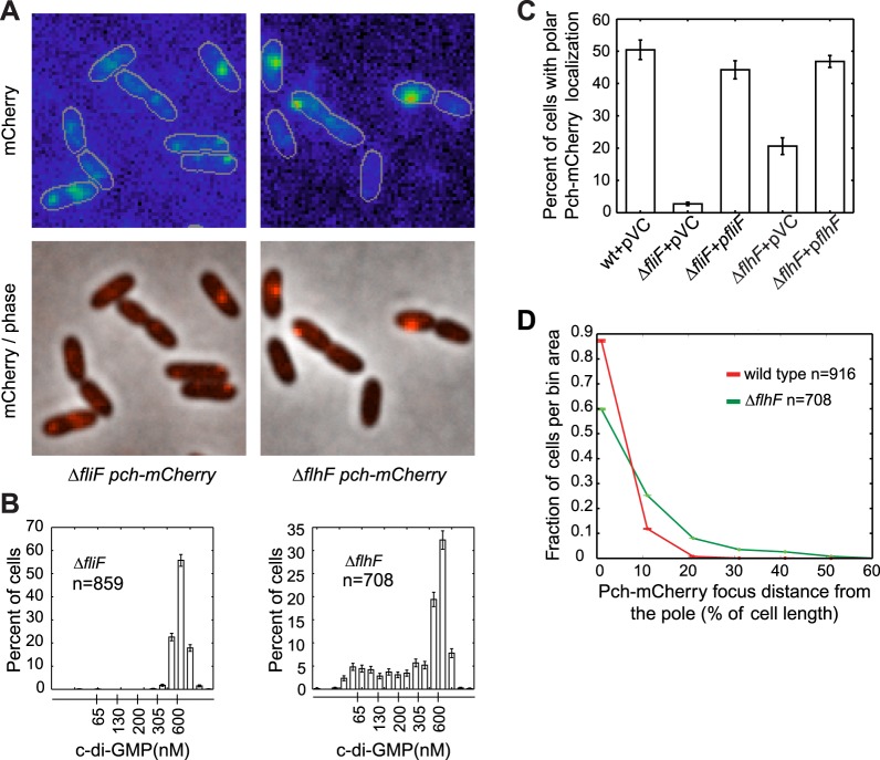
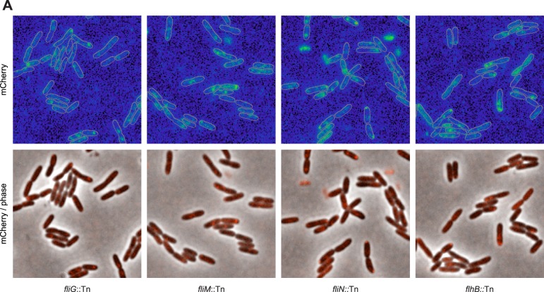
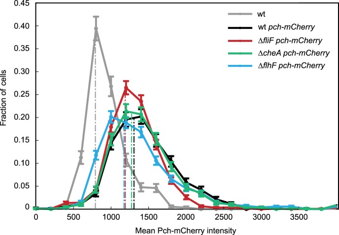
Figure 3. The flagellar apparatus is required for Pch polar localization and maintenance of low c-di-GMP.
(A) Representative images (10.8 × 10.8 microns) of Pch-mCherry localization in fliF and flhF deletions. Refer to the ‘Materials and methods’ section for a description of contrast settings for both image types. (B) Histograms of cellular c-di-GMP concentrations in fliF and flhF deletions. Error bars represent counting error. The mean percentage of ΔfliF cells with less than 200 nM c-di-GMP from three biological replicates is 0.1% (standard deviation of 0.1%). The mean percentage of ΔflhF cells with less than 200 nM c-di-GMP from three biological replicates is 26.6% (standard deviation of 5.2%). (C) Quantification of the mean percentage of cells exhibiting polar localization of Pch-mCherry in ΔfliF and ΔflhF backgrounds from four biological replicates. Error bars represent the standard deviation. Strains contain empty vector (pVC) or a complementing plasmid. Quantification was performed as described in the ‘Materials and methods’. (D) A representative histogram of Pch-mCherry focus distance to the nearest cell pole in wild type and flhF deletion strains. Error bars depict counting error.


