
Figure 2.
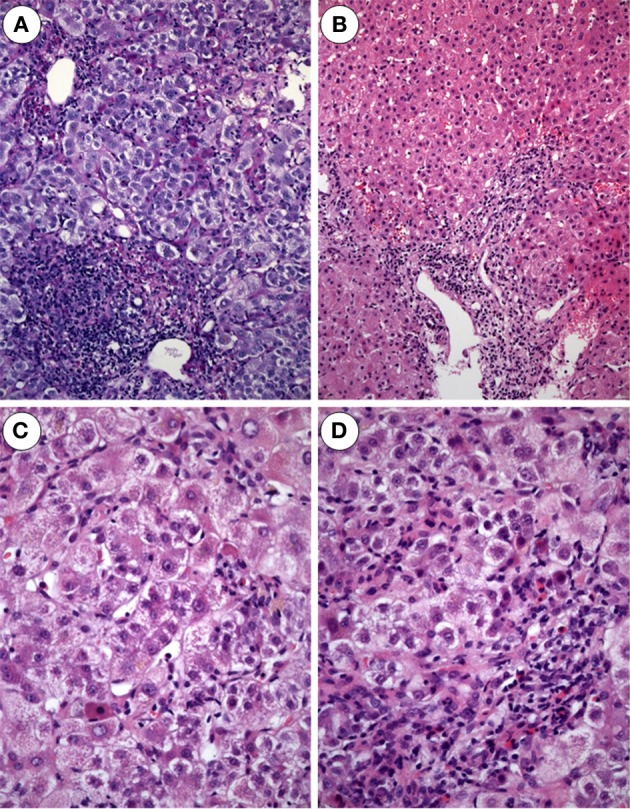
Acute hepatitis E. (A) Expanded portal tract with dense inflammatory infiltrates mostly lymphocytes. Bile ducts display mild accompanying cholangitis (H and E × 100). (B) Acute hepatitis E with enlarged portal tract densely infiltrated by lymphocytes and some PMN leukocytes as well as some spotty necroses in the lobule (H and E × 80). (C) Acute hepatitis E with areas of spotty necrosis, aptotic bodies and infiltrates of lymphocytes, Kupffer cells and few polymorphnuclear leukocytes (H and E m× 240). (D) Biopsy from a patient with acute hepatitis E: the lobule shows foci of spotty necrosis, ballooning of hepatocytes and infiltrates with lymphocytes and polymorphnuclear leukocytes (H and E × 240)