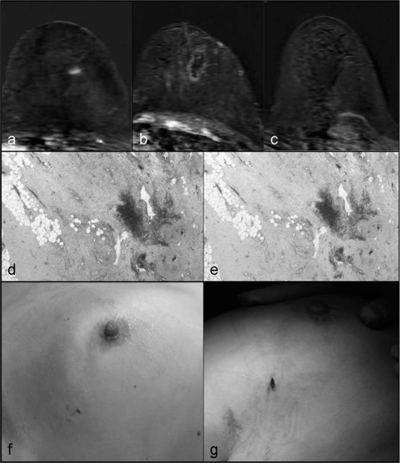
Fig. 1.
Radiofrequency ablation, histological examination, and cosmetic evaluation. a Axial T1-weighted magnetic resonance imaging (MRI) showing an enhancing lesion in the upper-outer left breast; b ablation volume after percutaneous ablation; c absence of enhancement at a delayed MRI; d hemorrhagic/necrotic areas (hematoxylin and eosin) without viable epithelial cells; e NADH diaphorase; f excellent cosmetic results at Time 0 after percutaneous ablation, g at Time 1 prior to surgical excision with evidence of mild pigmentation of the skin.

