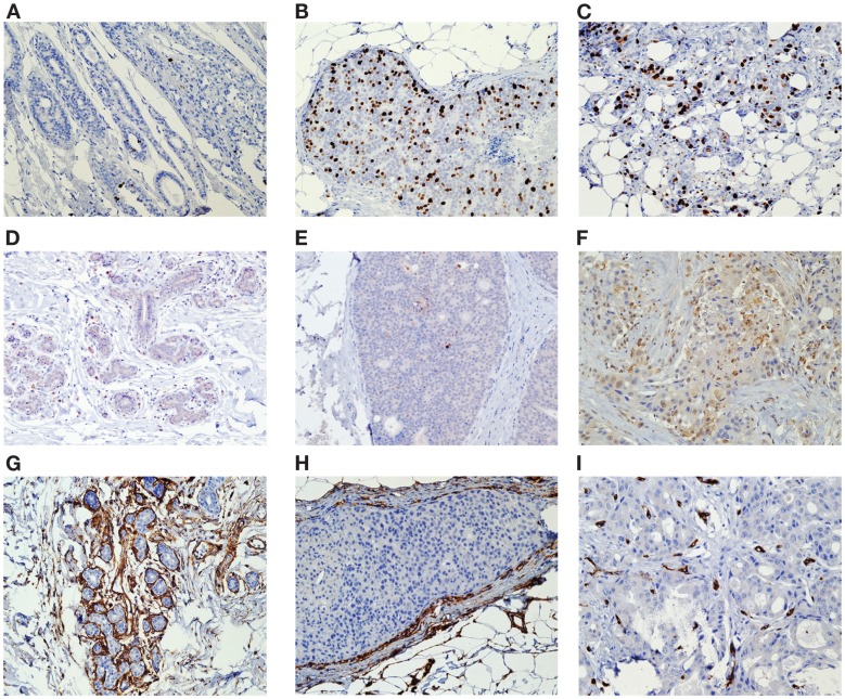
Figure 1.
Tissue was stained for specific markers of cell proliferation (Ki-67), apoptosis (casp-3), and angiogenesis (CD34) using the respective antibodies. Separate IHC analyses were performed on the benign and malignant (DCIS and invasive) breast cell components Ki-67 Histology of biopsy benign (A), DCIS (B), and invasive tissue (C) Caspase-3 Histology of biopsy benign (D), DCIS (E), and invasive tissue (F) CD34 Histology of biopsy benign (G), DCIS (H), and invasive tissue (I).

