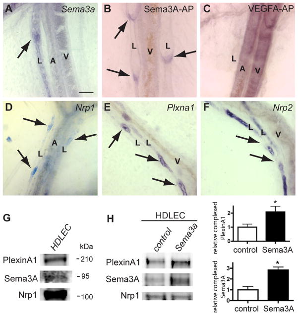
Figure 1. Sema3A expression and binding to lymphatic valves expressing Nrp1 and PlexinA1.
A, In situ hybridization with Sema3a antisense riboprobe on P0 mesenteric vessels. Note specific labeling of lymphatic vessels (L) including valve-forming areas (arrow) but not arteries (A) or veins (V). B and C, Sema3A-AP (B) and VEGFA-AP (C) fusion protein binding to whole-mount mesenteries from wild-type mice at P0. Sema3A-AP binds to lymphatic valves (B, arrows). VEGFA-AP binds to arteries and veins but not to lymphatics (C). D through F, In situ hybridization of whole-mount mesenteries at P0 with the indicated antisense riboprobes. Nrp1 and Plxna1 label valve-forming areas of lymphatic vessels (D and E, arrows). Note strong Nrp2 staining in lymphatic vessels but absence of signal in valve-forming areas (arrows, F). G, HDLEC protein extracts subjected to sodium dodecyl sulfate polyacrylamide gel electrophoresis and Western blots were incubated with the indicated antibodies. HDLEC express Nrp1, PlexinA1, and Sema3A. H, HDLEC were serum-starved and stimulated with Sema3A for 15 minutes. Nrp1 was immunoprecipitated from cultures, and Western blots were probed with indicated antibodies. Quantification of 3 independent experiments shows that Sema3A treatment enhances Nrp1/PlexinA1 complex formation. Bars represent SEM. *P<0.05 (t test). Scale bars: 50 μm.

