
Figure 7.
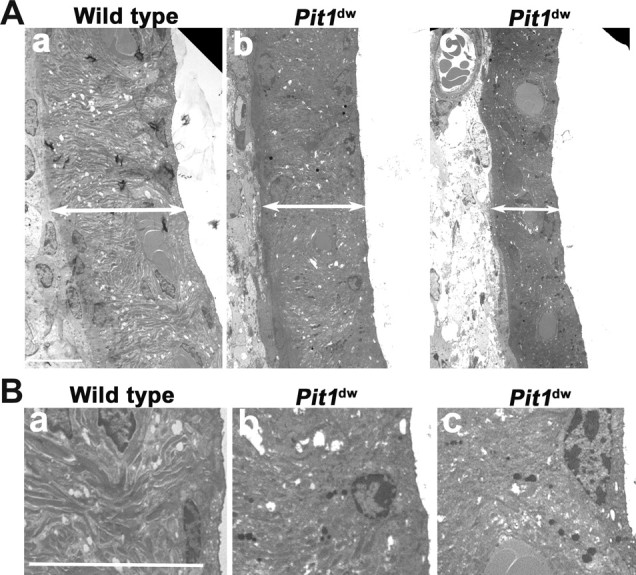
Stria vascularis pathology in adult Pit1 mutants. A, Stria vascularis prepared from P42 wild-type mice (a) and mutant mice (b, c) were analyzed by TEM. Double-headed arrow bars define the width of the stria vascularis. B, Lipofuscin granules are more abundant in the intermediate cells of Pit1dw mutant mice, suggesting that the mutant cells are exhibiting signs of aging. The ultrastructure of wild-type (a) stria vascularis animals was compared with Pit1dw mutants (b, c). Lipofuscin-like granules are more abundant in the mutants. Sections shown are from the basal turns of the cochlea, which were more prominently affected than the apex. Scale bars: 10 μm.