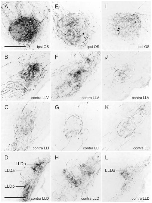
Figure 4.
A–D: Fiber and terminal labeling in the left (ipsilateral) OS (A) and right (contralateral) LLV (B), LLIr (C), and LLDp (D) following an injection of BDA into medial NA shown in Fig. 3A,B. E–H: Fiber and terminal labeling in the left (ipsilateral) OS (E) and right (contralateral) LLV (F), LLIr (G), and LLDp (H) following an injection of BDA into lateral NA shown in Fig. 3C. I–L: Fiber and terminal labeling in the left (ipsilateral) OS (I) and right (contralateral) LLV (J), LLIr (K), and LLDa (L) following an injection of BDA into mid-NL shown in Fig. 3E. Scale bars = 200 μm for A–K; 400 μm for D,H,L.

