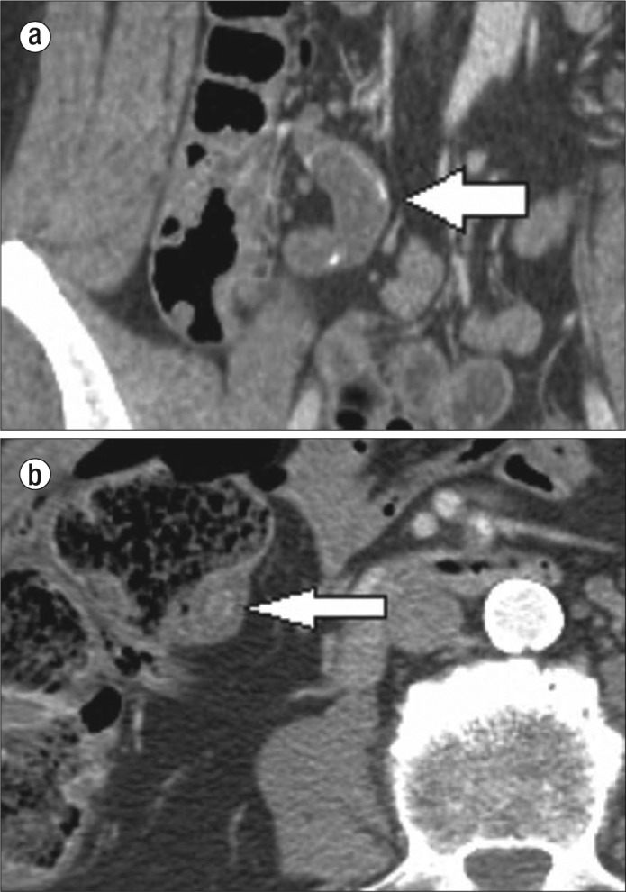
Figure 1.

Mucocele of the appendix. (a) Coronal CT image of the right lower quadrant in patient 1 shows a dilated appendix with thin calcification in the wall (white arrow). (b) Axial CT image of the right lower quadrant in patient 2 shows a dilated appendix with an appendicolith.
