
Fig. 13.
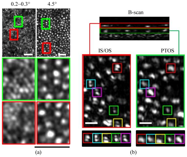
Photoreceptor mosaics extracted from AO-OCT volumes acquired on myopic subjects. (a) En face projections confined to the photoreceptor layer is shown for volumes acquired at 0.2°~0.3° and 4.5° retinal eccentricity in the left and right columns, respectively. Scale bars are 25μm (b) OCT B-scan is shown at the top with corresponding en face images of (left) IS/OS and (right) PTOS reflections below at 6° retinal eccentricity. Inset shows individual cones that exhibit an IS/OS reflection characteristic of a TEM10–like mode. Superimposed boxes are color coded and 7.8 μm in width. Scale bars are 10 μm.