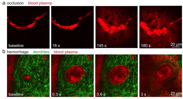
Figure 1. Femtosecond laser ablation can be used to generate experimental models of ischemic and hemorrhagic microstroke.
a) Time-lapse, two-photon imaging of fluorescently labeled blood plasma (red) during the formation of an occlusion in a targeted sub-surface brain arteriole. Irradiation by a series of tightly-focused femtosecond laser pulses is used to injure the endothelium of the targeted vessel and trigger clotting. b) Hemorrhage from a penetrating arteriole generated by irradiation with a single high-energy laser pulse. Neuronal processes (green) are displaced by the expanding hematoma while blood plasma (red) pushes into the brain parenchyma. Red blood cells are visualized as dark volumes within the fluorescent blood plasma. Time stamps indicate the time after the first laser irradiation.

