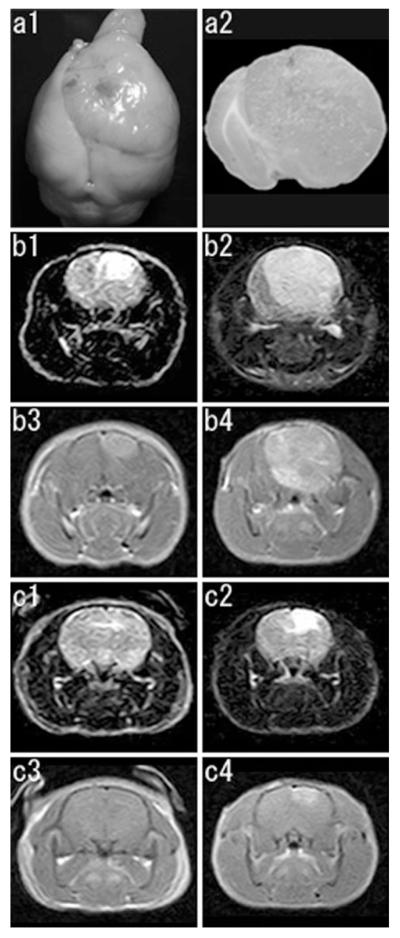
Fig. 1.
a Macroscopic features of a PDGF-induced tumor (GBM, viral injection at P10, 42 days post injection, the same animal as presented in b). Dorsal view of the whole brain (a1),and a coronal brain slice at the level of the viral injection (a2). A large tumor is noted with remarkable midline shift. b, c Serial MRI studies of PDGF-induced tumors. Representative coronal images are presented (b1, b2, c1, c2, FLAIR images; b3, b4, c3, c4, T1 weighted images with gadolinium enhancement). b This animal shows development of a tumor at 34 dpi (b1, b3) and its rapid progression at 48 dpi (b2, b4). c This animal shows no tumor formation at 34 dpi (c1, c3), but development of a tumor at 48 dpi (c2, c4)

