
Fig. 3.
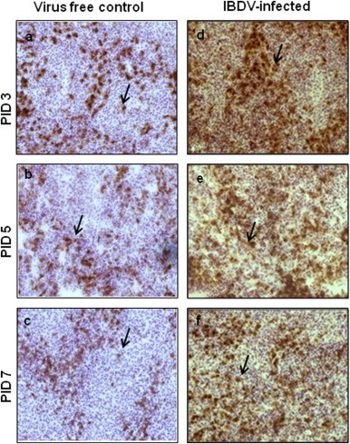
Infiltration of CD8+ T cells in the spleen. Three-week-old chickens were inoculated with IBDV and frozen sections of spleen tissues were prepared. At PIDs 3, 5 and 7 spleen sections from virus free chickens (a, b, and c) and IBDV-infected chickens (d, e, and f) were examined (40×) for the presence of CD8+ T cells by immunohistochemistry using anti-chicken CD8+ mAb. Brown color shown by arrow indicates the positive staining.