
Fig. 7.
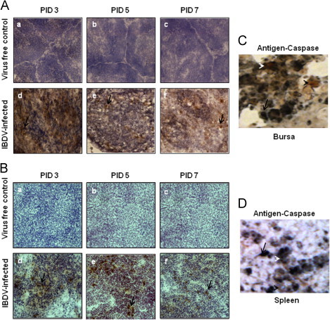
Detection of cleaved caspase-3 in bursa and spleen (A-B). Chickens were inoculated with IBDV; bursa and spleen tissues were collected. At 3, 5 and 7 PID, bursa and spleen sections from virus free chickens (a, b and c) and IBDV-infected chickens (d, e, and f) were examined (40X) for the presence of activated cleaved caspase-3 by immunohistochemistry using antihuman cleaved caspase-3mAb. Development of brown color shown by arrow indicates positive staining for cleaved caspase-3. Bursal and splenic tissue sections (C-D) were double stained by immunohistochemistry using cleaved caspase-3 and anti-IBDV R63 monoclonal antibodies. Blue staining shown by arrow represents IBDV antigen positive cells and brown staining represented by black arrow head shows caspase positive cells.