
Table 1.
Detection and quantification of CD8+ T cells and cytotoxic T cells mediators: Fas, FasL, perforin and cleaved caspase-3 by immunohistochemistry in IBDV-infected chickens.
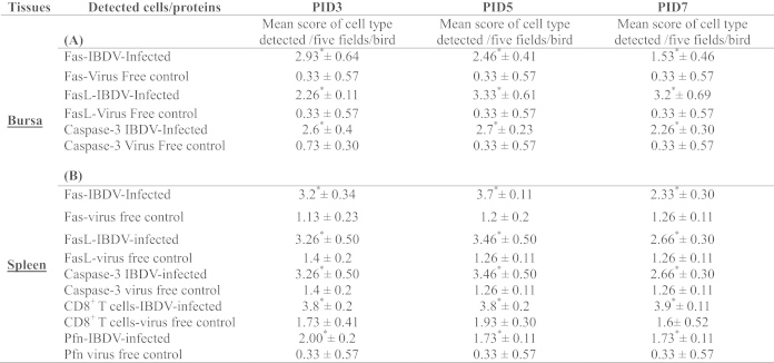 |
Foot notes. Three weeks old SPF chickens were inoculated with 104EID50 of cIBDV. Bursa and spleen tissues were collected at PIDs 3, 5 and 7. Bursal and spleens sections from virus-free chickens and cIBDV-infected chickens were examined for the detection of CD8+ T cells and cytotoxic T cells mediators: Fas, FasL, Perforin and cleaved caspase-3 by immunocytochemistry. Each immunostained section was examined (20×) and given positive cells count based on each cell type (Scores of positive cells/field: 1=1–25%; 2=26–50%; 3=51–75%; 4=76–100%). Student's t-test was used to detect significant differences between virus free control and IBDV-infected chickens. The values represent the mean ±SEM of 5 fields/bursa and/or spleen/chicken on designated PID (3, 5 and 7). * indicates statistically significant differences between virus free control and IBDV-infected groups (p<0.05).