
Fig. 4.
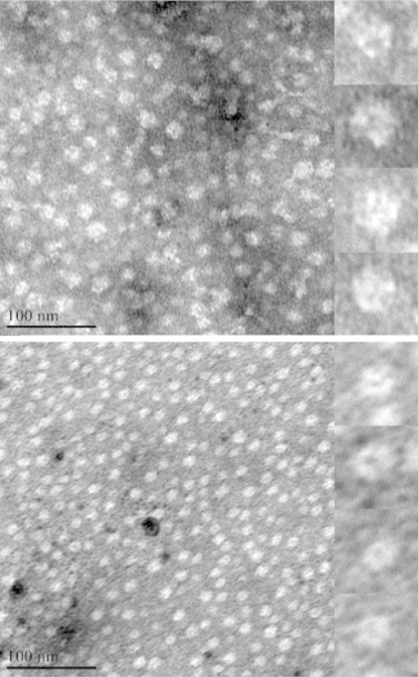
Negative stain TEM analysis of globular TbHK1 hexamers. Samples of WT TbHK1 (upper) and C445A variant TbHK1 (lower) were analyzed using a JEOL JEM 1200 EX transmission electron microscope after negative staining. Inset contains enlargement of four representative protein particles. Scale bar = 100 nm.