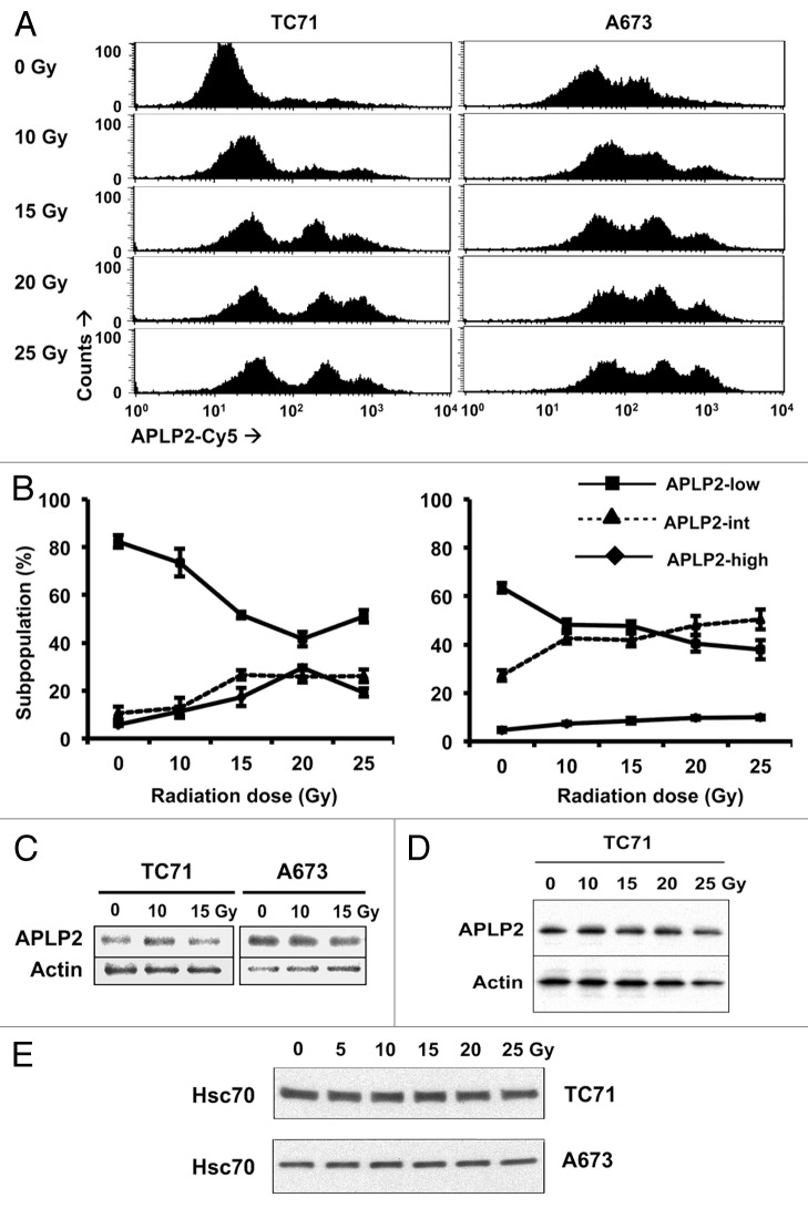
Figure 2. APLP2 is expressed on the surface of EWS cells and can be redistributed by ionizing radiation. (A-E) Radiation shifts EWS cells toward subpopulations with high levels of surface-exposed APLP2, but does not increase total APLP2 expression. EWS cells were irradiated with the indicated dose of γ rays and cultured for 24 h prior to analysis. (A) Representative histograms of surface APLP2 expression on TC71 (left) and A673 (right) cells 24 h post-irradiation as determined by flow cytometry. (B) The mean frequencies of EWS cells with APLP2low (box), APLP2int (triangle) and APLP2high (diamond) phenotypes are shown with respect to radiation intensity. Error bars denote SD (n = 6 experimental replicates within 1 experiment that yielded results representative of several experiments (2 experiments including both TC71 and A673, 2 with A673 but not TC71, and 3 with TC71 but not A673). (C–E) Irradiated TC71 and A673 cells were lysed, and the lysate supernatants were subjected to immunoblotting to determine total expression levels of APLP2 (C and D). Actin levels were monitored to ensure equal lane loading in C and D. Separate experiments were performed to demonstrate that there is no decrease in the expression of another loading control, HSC70, in TC71 and A673 cells exposed to 25 Gy (E). Immunoblotting data are representative of n = 2-3 independent experiments yielding similar results.

An official website of the United States government
Here's how you know
Official websites use .gov
A
.gov website belongs to an official
government organization in the United States.
Secure .gov websites use HTTPS
A lock (
) or https:// means you've safely
connected to the .gov website. Share sensitive
information only on official, secure websites.
