
Figure 5. The follow-up scans of trauma-exposed victims with PTSD showed decreased FA in left insula WM compared with their baseline DTI scans.
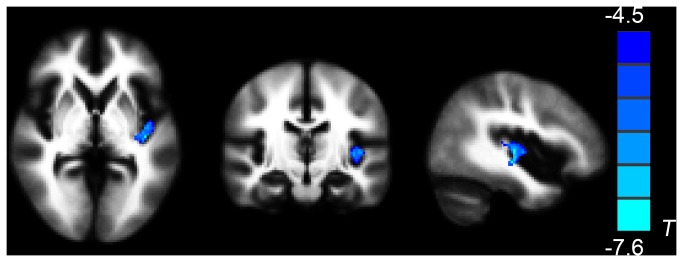
(paired t-sample t-test, P < 0.05, AlphaSim-corrected).
Cold colors indicate decreases in FA. The left part of the figure represents the participant’s right side.
PTSD = posttraumatic stress disorder; FA = fractional anisotropy.