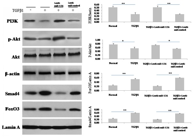
Figure 5. miR-126 activated the PI3K/Akt and inhibited the FoxO3/Smad4 signalling pathways in TGFβ1-induced EPCs.

After the treatment with TGFβ1 (5 ng/mL) for 7 days, the protein in EPCs were prepared. Immunoblotting assays were performed using specific antibodies against PI3K, phosphor-Akt, Akt, FoxO3, and Smad4. β-actin and lamin A were used as internal controls for total cells and the nuclear proteins assay separately. The relative protein levels of these proteins were determined by densitometry analysis (n = 3). Data are shown as mean ± S.D. values. * and ** represent P < 0.05 and P < 0.01 with an LSD t-test, respectively. The number of observations (n) represents the number of independent cell preparations.
