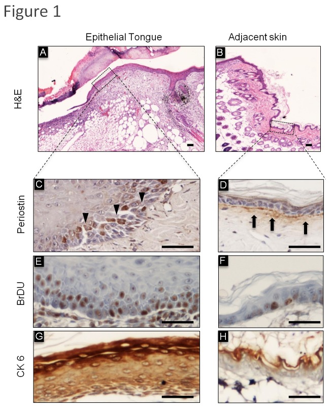
Figure 1. Expression of Periostin and CK6 during cutaneous wound healing.
H&E: Representative histological sections of cutaneous incisional wounds. (A) Morphology of the wounded site shows a thin edge of epithelial cells migrating across the wound bed, termed the epithelial tongue and (B) intact and normal skin adjacent to the wounded site were stained with Hematoxylin and Eosin (H&E). (C) Epithelial cells at the epithelial tongue express intracellular Periostin. (D) In normal adjacent skin, Periostin is in the connective tissue at the basal lamina, which is juxtaposed to the epithelial basal layer. (E) Note that basal and parabasal layers of the epithelial tongue have a large number of proliferating BrDU positive cells. (F) As expected, the epithelial basal layer of adjacent skin has few proliferating cells. (G) Upregulation of the epithelia stress/tension marker CK6 is depicted in the epithelial tongue compared to normal adjacent skin observed in (H). Scale bars represent 50 μm.

