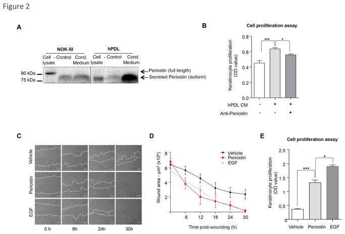
Figure 2. Periostin-driven migration and proliferation.
(A) Total cell lysates and conditioned medium (cond. medium) from NOK-SI and hPDL cells were blotted for Periostin. New cell culture medium supplemented with 10% FBS was used as a negative control (control). Intracellular Periostin is detected in epithelial cell lysate. However, conditioned medium from NOK-SI shows that keratinocytes did not secrete Periostin, as the same band was observed in the negative control media. hPDL cells have low levels of the intracellular Periostin isoform as observed in the cell lysate. Increased levels of secreted Periostin were found in the conditioned medium from hPDL. (B) hPDL conditioned medium induces keratinocyte proliferation compared to vehicle alone (***p<0.001), which is reduced upon administration of anti-Periostin antibody (*p<0.05). (C) Representative pictures of NOK-SI migration following treatment with recombinant Periostin (50 ng/ml), EGF (100 ng/ml) as the positive control, or vehicle. Scale bars represent 50 μm. (D) Graphic represents the quantification of the wound areas at indicated times (n=4; mean ± S.E.M). (E) Periostin enhances proliferation of keratinocytes compared to vehicle treated cells (***p<0.001). EGF treatment was used as positive control (*p<0.05) (n=6; mean ± S.E.M).

