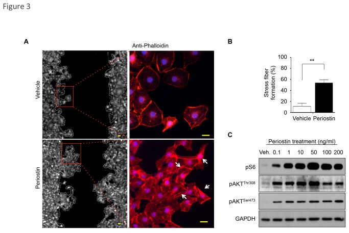
Figure 3. F-actin polarization and PI3K/mTOR signaling activation by Periostin-induced epithelial cell migration.
(A) Phalloidin detection shows cells with polarized F-actin (white arrow) following treatment with recombinant Periostin compared to vehicle control. Scale bars represent 10 μm. (B) Graphic represents percentage of cells with stress fiber formation (polarized F-actin) after periostin or vehicle stimuli. Results were determined by measuring fields using independent triplicates (**p<0.01) (C) Activation of PI3K and mTOR signaling is triggered by Periostin treatment in a dose-dependent manner, as detected by phosphorylated AKT at Threonine 308 (pAKTThr308) and Serine 473 (pAKTSer473) and phosphorylated S6 (pS6). Note that 50 ng/ml of Periostin is the optimal concentration for PI3K activation. GAPDH was used as a loading control.

