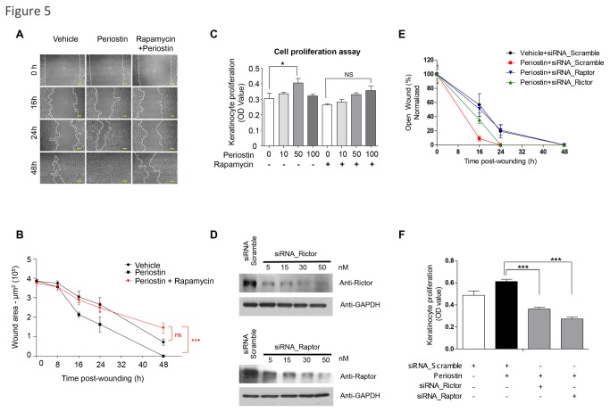
Figure 5. Periostin-driven cellular migration requires mTOR signaling.
(A) Representative pictures of the NOK-SI cell scratch assay following treatment with vehicle, recombinant Periostin (50 ng/ml), and rapamycin (50 nM). Scale bars represent 50 μm. (B) Quantitative analysis of open-wounded area over time (n=4; mean ± S.E.M.). Note that rapamycin abrogates the Periostin migratory activity of epithelial cells (***p<0.001). (C) Proliferation assay using keratinocytes treated with rapamycin and/or Periostin. Note that Periostin alone induced significant cellular proliferation at 50 ng/ml (*p<0.05). Treatment with rapamycin blocked periostin-induced cell proliferation (ns p>0.05). (D) Representative immunoblot depicting knockdown of Raptor and Rictor after siRNA treatment. Scramble siRNA oligonucleotides sequences were used as controls. GAPDH was used as loading controls. (E) Graphic shows the quantitative analyses of open-wounded areas using NOK-SI cells over time (n=4; mean ± S.E.M.). Note that siRNA targeting Raptor abrogates Periostin-induced cellular migratory resulting on complete wound closure by 48 hours (**p<0.05). siRNA targeting Rictor did not change the Periostin induced accelerated cellular migration resulting on wound closure by 24 hours (ns p>0.05). (F) Proliferation assay using NOK-SI cells treated with siRNA for Raptor, Rictor, or siRNA scramble, and/or Periostin. Note that Periostin induced significant cellular proliferation (*p<0.05). Treatment with siRNA for Raptor or Rictor resulted in disruption of Periostin induced cellular proliferation (***p<0.001).

