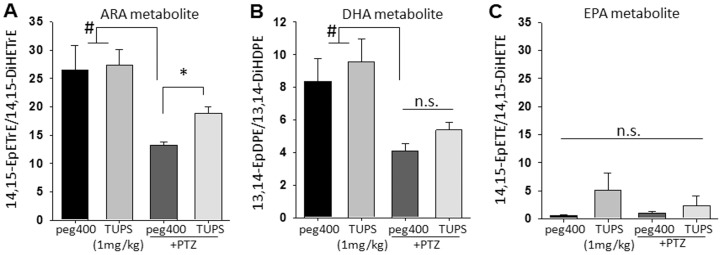
Figure 5. Brain oxylipins display decrease in response to seizure and are elevated by sEHI.
(A–C) Bar graph of ratios of brain epoxy to dihydroxy-FAs in mice receiving vehicle (n = 6, black bars), sEHI (n = 6, gray bars), vehicle+PTZ (n = 8, dark gray bars) and sEHI+PTZ (n = 8, light gray bars). Inhibitors were administered 1 h prior to sampling, PTZ treated animals were sampled up immediately following tonic phase of the seizure. PTZ treatment resulted in a significant decrease in the levels of bioactive regioisomers 14,15-EET and 13,14- EpDPE (Kruskal-Wallis One Way ANOVA on Ranks followed by Dunn's multiple comparison, control vs PTZ, # p≤0.05). Although in the absence of seizures sEHI did not lead to changes in the brain levels of 14,15- EET and 13,14- EpDPE, the decrease mediated by seizures was recovered by inhibition of sEH for the ARA metabolite 14,15-EET (*p = 0.001) but not for the DHA metabolite 13,14-EpDPE (p = 0.061). There were no significant changes in the levels of EPA derived epoxy fatty acids among groups.

