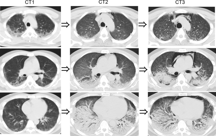
Figure 2.
CT on admission showed attenuation adjacent to the pleura in the upper and middle fields of both lungs (CT1). After steroid pulse therapy, the CT showed that the opacities decreased in the upper field but a denser area appeared in the lower field. Slight mediastinal emphysema also appeared (CT2). CT when the lung had lost a lot of function showed centrilobular nodules in the upper and middle lobes, and the volume of the lower lobe decreased due to traction bronchiectasis. Furthermore, left and right pleural effusions appeared (CT3).

