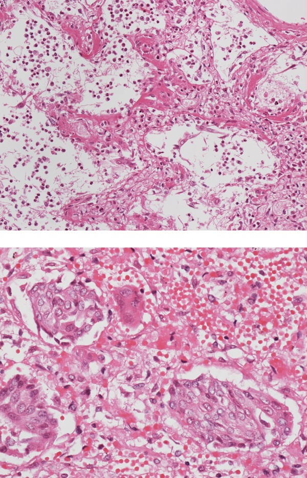
Figure 5.

Pathological examination revealed diffuse alveolar damage in the entire lung. Oedema, haemorrhage, proliferation of type II pneumocytes, thickening of the vascular wall and centrilobularly distributed squamous metaplasia were also observed. In some areas, the hyaline membrane lining the alveolar duct was seen (A). Notably, squamous metaplasia was more prominent than that observed in conventional acute interstitial pneumonia (B).
