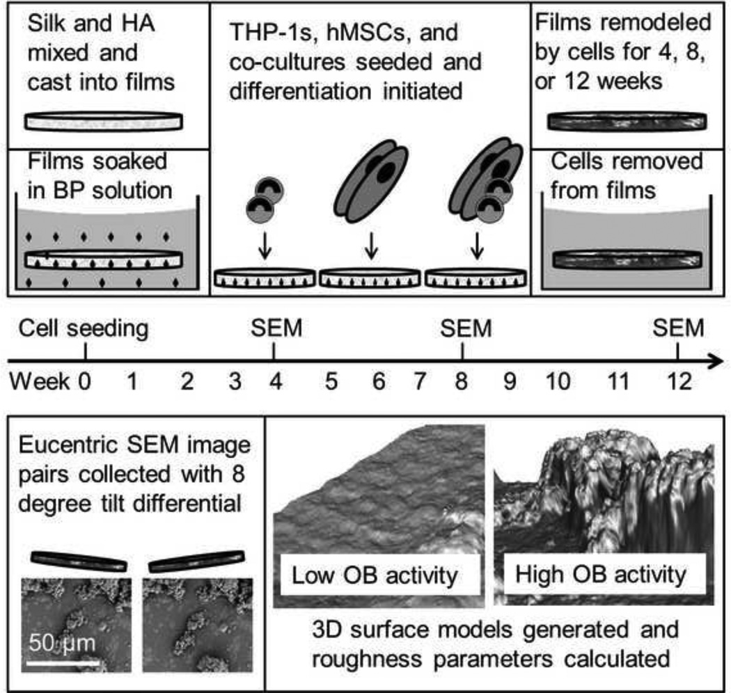
Figure 1. Schematic of 12 week studies.
Top: Films were cast from a dispersion of HA in aqueous silk solution. Following drying and water annealing, films were soaked in solutions of clodronate or alendronate which bound to the HA. Following autoclaving, films were seeded with hMSCs, THP-1s, or a co-culture of the two cell types in equal number. Differentiation was then initiated, and films were remodeled by cells for 4, 8, or 12 weeks. For surface metrology analysis cells were removed from films by soaking in water overnight at 4°C and films were dried and sputter coated. Bottom: Eucentric SEM images were taken with an 8 degree difference in tilt angle. 3D surface models were generated and roughness parameters were calculated. Example surfaces from low and high osteoblast activity (approximately 600 µm2) are shown.

