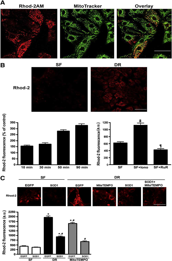
Fig. 2.
A: Co-localization of rhod-2 and MitoTracker fluorescence in LLC-PK1 cells. B: Time course of rhod-2 fluorescence following 1 h ATP depletion and varying recovery times indicates progressive accumulation of mitochondrial Ca2+. Ca2+ ionophore, ionomycin increases mitochondrial Ca2+, which is blocked by inhibiting mitochondrial Ca2+ uptake with ruthenium red. ¶, P < 0.01 Ruthenium vs. serum free (SF); ‡, P < 0.001 Ionomycin vs. SF. C: Representative images (top panel) and summary histograms of rhod-2 fluorescence intensity (bottom bar graph) in LLC-EGEP and LLC-SOD1 cells following 1h-1h ATP depletion- recovery. Results are means ± SEM, with N=80–100 cells from 4 different dishes. *, P < 0.05 vs. SF; #, P < 0.05 vs. EGFP-DR. Scale bar = 25 µm for 1A, 40 µm for 1B and 1C.

