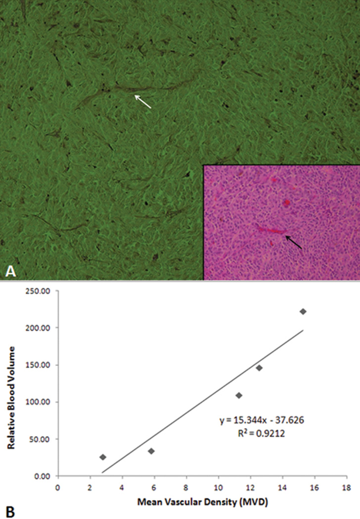
Figure 4.
Tumor mean vascular density (MVD) correlates with sonographic tumor blood volume. (A) Histologic section of the melanoma stained with periodic acid-Schiff (PAS) without hematoxylin is used to determine mean vascular density. A blood vessel in the PAS stained section (white arrow) correlates with the section stained with hematoxylin and eosin (inset, black arrow). (PAS ×100; inset hematoxylin and eosin, 100×) (B) The relative blood volume obtained from the ultrasound correlates with the MVD measured from the histologic sections (R2=0.9212).

