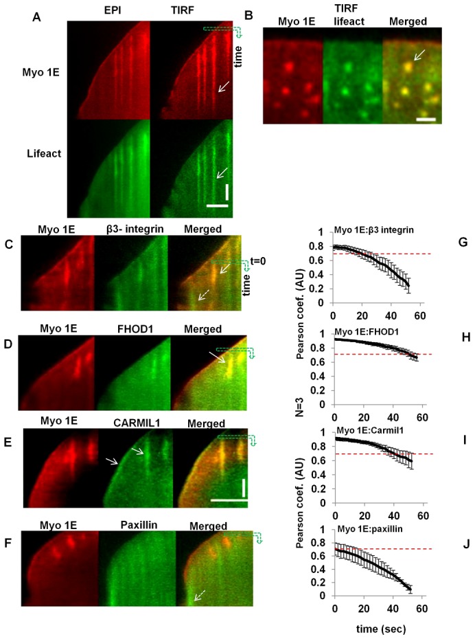
Fig. 2. β3-integrin, FHOD and CARMIL co-localize with Myosin 1E in actin rich early adhesion.

Paxillin shows partial overlap. (A) Simultaneous observation (kymograph) of Myosin 1E and actin (lifeact) in EPI and TIRF channels (supplementary material Movie 2). Time is along vertical axis along the green arrow, length is along horizontal axis. Bar 5 µm. (B) Co-localization of Myosin 1E with actin rich spots (white arrow). Bar 2 µm. Comparative co-localization of (C) EGFP-β3-integrin, (D) FHOD1, (E) CARMIL1a and (F) Paxillin with mApple-Myosin 1E at the tip and early adhesion structures in spreading lamellipodia (by kymograph, time as vertical and length as horizontal axis). Bar 5 µm, C–F is in same scale, time bar 30 sec. (G–J) Quantification of such overlap of Myosin 1E with β3-integrin/FHOD1/CARMIL1/paxillin by Pearson coefficients (starting point or zero sec is indicated in the kymograph as the starting point of downward green arrows in C–F) (associated supplementary material Movies 3–6).
