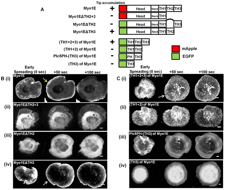
Fig. 4. Requirement of multiple domains of Myosin 1E in lamellipodial tip accumulation.
(A) Domain map of various myosin 1E deletion (C-terminal and N-terminal) mutants and summary of their lamellipodia tip accumulation during cell spreading process. (B) Edge accumulation of C-terminal deletions. (Bi) Myosin 1E (full length) at the tip of spreading lamellipodia (yellow arrow). No such accumulation in (Bii) Myo1EΔTH2+3 and (Biii) Myo1EΔTH2. Such accumulation was present in (Biv) Myo1EΔTH3 (white arrow). (C) Edge accumulation of N-terminal deletions. (Ci) (TH1+2+3) at the tip of spreading lamellipodia (white arrow). No such accumulation in (Cii) (TH1+2), (Ciii) PlcδPH-TH3 and (Civ) TH3. Bar 5 µm.

