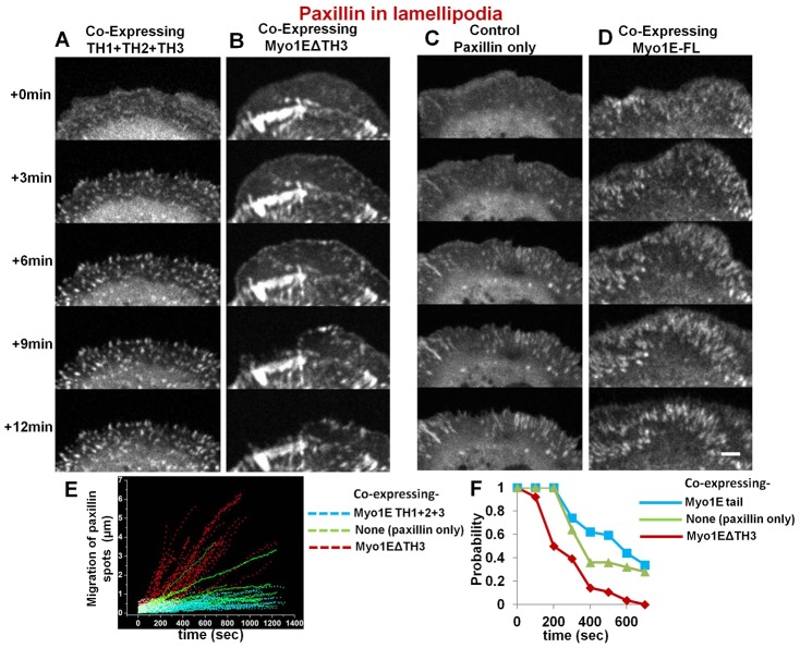
Fig. 6. Adhesion formation by paxillin fluorescence: between cells co-expressing full or deletion mutants of Myosin 1E and control cell expressing paxillin alone, showing dominant negative effects of deletion mutants.
Dot-like structures of expressed paxillin in lamellipodia of cells co-expressing (A) (Th1+2+3) or (B) Myo1EΔSH3. In (B) newly formed adhesions showed instability in lamellipodia (supplementary material Movie 13). Elongated paxillin structures in active lamellipodia denoting stable control cells expressing paxillin only (C) or co expressing full length Myosin 1E (D). Bar 5 µm. (E) Movement of nascent adhesions in dynamic lamellipodia, as observed by RFP-paxillin spots; observed in cells co-expressing Myo1EΔSH3/(Th1+2+3) and control cells expressing paxillin alone. (F) Probability of finding a paxillin spot within 1 µm radius from where it first appeared in dynamic lamellipodia with time. These nascent adhesions (paxillin-spots) were compared from three cells each (per construct) co-expressing Myo1EΔSH3, (TH1+2+3) or control cells expressing paxillin alone.

