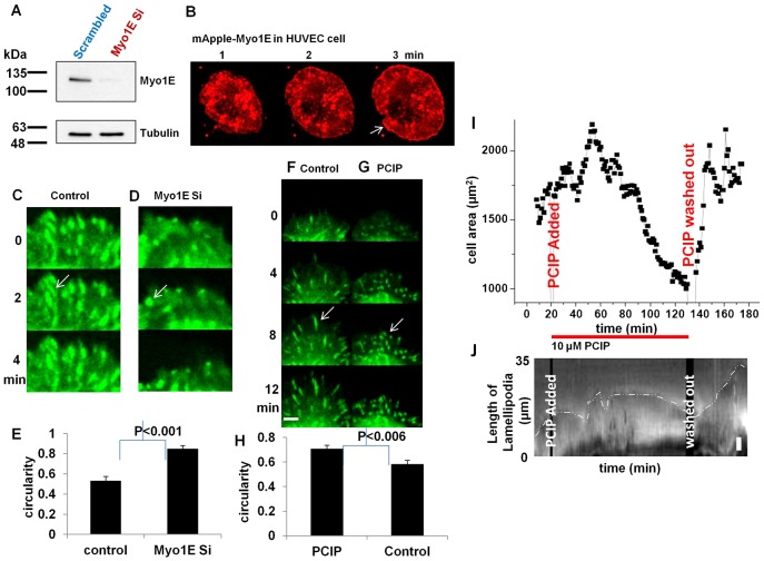
Fig. 7. Myosin 1E knockdown has destabilizing effect on cell–matrix adhesions. Similar distablization observed in Myosin 1 inhibitor treated cell.
(A) HUVEC cells expressing Myosin 1E, showing edge accumulation (arrow). (B) Knockdown of Myosin 1E in HUVEC cells, (C) Control and (D) Myosin 1E siRNA transfected HUVEC cells (after two consecutive transfections) show difference in circularity of paxillin staining at adhesions (white arrows, supplementary material Movie 14). (E) Myosin 1E siRNA transfected cells have significantly higher circularity than control cells. (F) Control and (G) PCIP treated REF 52-paxillin-GFP cells show difference in circularity of paxillin staining at adhesions (white arrows, supplementary material Movie 15). (H) PCIP treated cells have significantly higher circularity than control cells. (I) Cell area constriction of actively spreading RPTP cells upon addition of PCIP (supplementary material Movie 16). Washout led to re-spreading to same cell area. (J) Kymographic representation of constriction of lamellipodia the cell shown in (I) upon addition of PCIP. Washout led to re-spreading of lamellipodia. Bar 10 µm in A, 5 µm in J, 2 µm in the rest.

