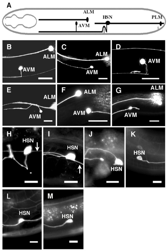
Fig. 1. Genetic interactions between unc-53 and unc-6 and between unc-53 and unc-5 affect the direction of axon protrusion.

(A) Schematic diagram of AVM and HSN axon outgrowth. Axon outgrowth is towards ventral UNC-6 sources. (B–G) Photomicrographs of L4 stage animals showing the direction of AVM axon protrusion from the cell body. Ventral is down and anterior is to the left. Scale bar: 20 µm. In the wild-type pattern, the AVM axon ventrally protrudes toward the ventral nerve cord (B). Loss of unc-6 function causes anterior protrusion (C). Axon protrusion in the unc-53 mutants is similar to that observed in wild-type animal. (D). In unc-53(n152);unc-6(ev400) double mutants, AVM axons frequently protrudes dorsally (E), posteriorly (F), or have short extra extensions (G). (H–M) Photomicrographs of L4 stage animals showing the direction of HSN axon protrusion form the cell body. Ventral is down and anterior is to the left. Arrow indicates the PLM axon. Scale bar: 10 µm. In the wild-type pattern, the HSN axon protrudes ventrally from the cell body. After reaching the ventral nerve chord the axon extends anteriorly and defasciculates from the cord to form synapses at the vulva (H). Loss of unc-6 function causes anterior axon protrusion (I). Although most unc-53(n152) mutants have the wild-type protrusion pattern, in unc-53(n152);unc-6(ev400) mutants, HSN axons frequently protrude dorsally (J), posteriorly (K), or have extra extensions (L). Although most unc-5(e53) mutants have the wild-type protrusion pattern, in unc-53(n152);unc-5(e53) mutants, HSN axons frequently protrude anteriorly (M). PLM is not shown in J, K, L, or M because axons often terminate early in unc-53(n152) mutants.
