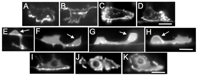
Fig. 5. EGL-20 inhibits anterior and posterior orientation of UNC-40 asymmetric localization and the formation of axons from these sites.

(A–D) HSN neurons expressing a functional UNC-40::GFP protein in the L3 larval stage. (E–H) HSN neurons in the L3–L4 larval transition stage expressing a marker to visualize the HSN neurons. Arrow points to HSN cell body. (I–K) HSN neurons expressing a functional MIG-10::GFP protein in the L3 larval stage. (A) In wild-type animals, the UNC-40::GFP protein is ventrally localized. (B) In unc-6 mutants, the UNC-40::GFP protein is uniformly dispersed around the periphery. (C and D) In egl-20 mutants, the UNC-40::GFP protein is localized ventrally as in wild-type animals or anteriorly (C) or posteriorly (D). (E) In wild-type animals the axon has a strong bias to ventrally protrude. (F) In unc-6 mutants, the axon has a bias to form anteriorly. (G and H) In egl-20 mutants the axon has a bias to form ventrally as in wild-type animals or anteriorly (G) or posteriorly (H). (I) In wild-type animals, the MIG-10::GFP protein is ventrally localized. (J,K) In unc-6 and egl-20 mutants, the MIG-10::GFP protein is uniformly dispersed around the periphery. Images are of collapsed stacks of optical sections. Ventral is down and anterior is to the left. Scale bar: 5 µm.
