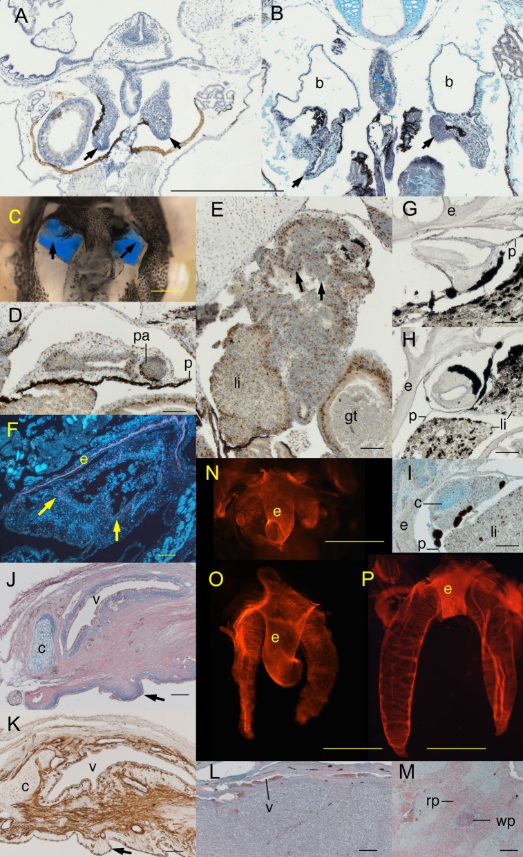
Fig. 2. Lung development in air-deprived (AD) and air-restored (AR) Xenopus.
(A,B) Frontal sections of AD tadpoles at (A) NF 46 showing normal lung development and (B) NF 49 showing expanded bronchi (b) and compressed, folded lungs (arrows). (C) Ventral view of a dissected AD NF 57 tadpole showing translucent stunted lungs against a blue background. (D,E) Frontal sections of an AD NF 57 tadpole showing (D) ventrally a compressed, thick walled bronchus with a pulmonary artery (pa) and small lumen anterior of the peritoneum (p) and (E) dorsally a compressed, folded lung abutting the liver (li) and gut tube (gt); arrows indicate incipient epithelial folds and brown is BrdU labeling. (F) Frontal section of a stunted AD NF 57 lung with fluorescently stained smooth muscle actin (red) and nuclei (white) showing little actin adjacent to the epithelium (arrows) and partial filling of the lumen by blood cells (the continuous actin layer is in the wall of the esophagus, e). (G–I) Frontal sections of AD NF 66+ frogs showing (G) ventrally a compressed bronchus, (H) dorsally the absence of a lung and the bronchus extending into the peritoneum-lined space normally occupied by the bronchial diverticulum, and (I) cartilage (c, blue) and BrdU labeling (brown) in the wall of a compressed bronchus. (J,K) Sagittal sections through the wall of a 19-month old AD lung showing a thick connective tissue layer containing cartilage (c), veins, and bands of actin fibers (pink in J and brown in K), and large incipient epithelial folds (arrows) protruding into the lumen (lower side). (L) A tissue-filled lung showing a thin wall, vein (v) and lumen filled with blood cells. (M) A section through the spleen of an adult frog showing the red (rp) and white pulp (wp). (N–P) Dorsal views of whole-mounted actin-stained lungs and esophagus (e) of (N) an AD NF 51 tadpole with weakly defined, closely spaced actin bands, (O) an AR NF 53 tadpole 9 days after air restoration with actin bundles more closely spaced than in untreated NF 52 lungs, and (P) an AR NF 59 tadpole 4 days after with widely spaced actin bundles but not the lateral subdivisions seen in untreated NF 59 lungs. Anterior is up for frontal views A–I and left for sagittal sections J,K. Scale bars are 0.5 mm for A,B; 1 mm for C, N and O; 0.1 mm for D–M; and 2 mm for P.

