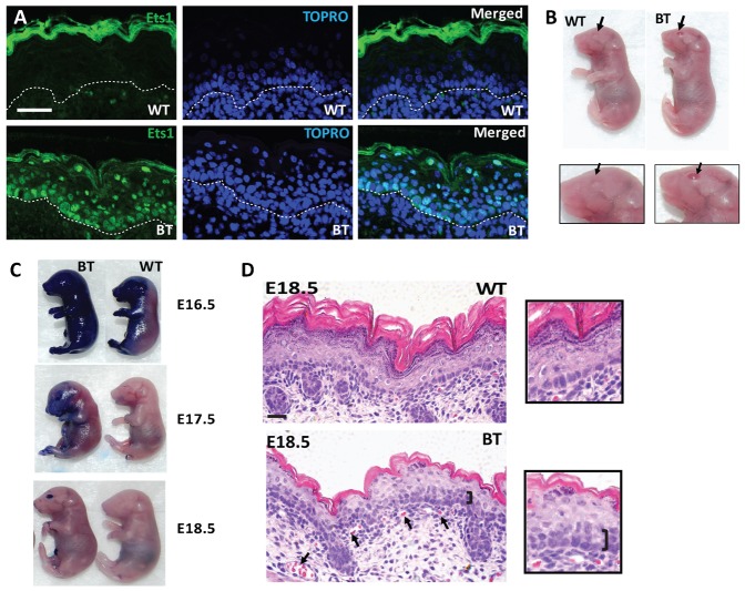
Fig. 1. Effects of over-expression of Ets1 in the basal layer of the skin.
(A) Immunofluorescent staining of E18.5 wild-type and K5-Ets1 BT skin for Ets1 (green staining). Sections were counterstained with TOPRO-3 to mark the nuclei (blue). Scale bars are 37.5 µm. (B) Embryonic E18.5 K5-Ets1 BT mice in which the Ets1 transgene was induced prenatally exhibit an eye-open-at-birth phenotype (arrows point to closed or open eyes in the wild-type and K5-Ets1 BT pups), but grossly normal skin. (C) K5-Ets1 BT embryos exhibit delayed skin barrier acquisition at E16.5 and E17.5. However, at E18.5, both wild-type and BT embryos show similar skin barrier function (note that the open-eye phenotype of BT embryos results in a blue-stained eye). (D) Hematoxylin and eosin (H&E) staining of E18.5 embryonic wild-type and BT skin demonstrates a second layer of basal-like epithelial cells (black bracket), impaired differentiation of suprabasal keratinocytes and increased angiogenesis (black arrows). Scale bars are 37.5 µm. Inset shows a higher magnification view of the skin.

