
Fig. 3. Phenotype of embryos injected with TALEN mRNAs.
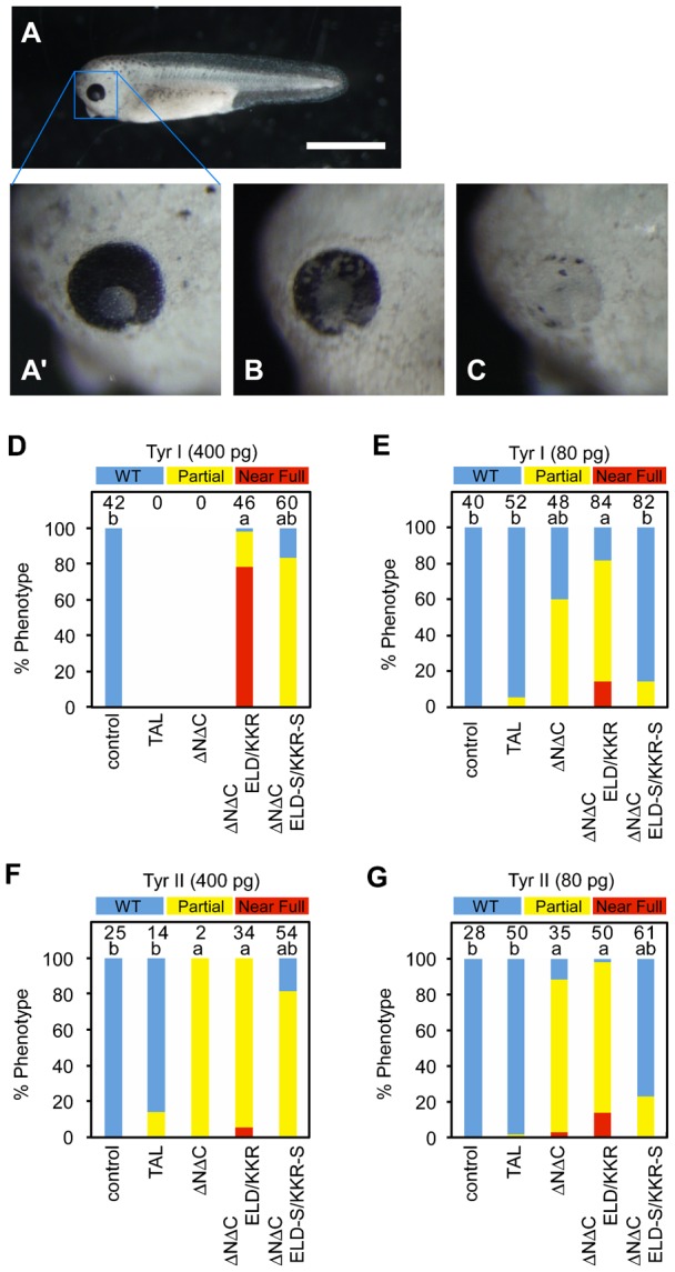
(A) A wild-type embryo at NF-stage 35/36. (A′) A higher magnification of A (WT). (B) An eye with less than 50% pigmentation loss in the retina (Partial). (C) An eye with more than 50% pigmentation loss in the retina (Near Full). (D–G) Percentages of wild-type eyes (blue), eyes with a partial loss (yellow), and eyes with a severe loss (red) of pigmentation in the retina of NF-stage 35/36 embryos injected with TAL, ΔNΔC, ΔNΔC-ELD/KKR or ΔNΔC-ELD-S/KKR-S mRNAs. The embryos were injected with 400 pg, 80 pg or 0 pg (control) of TALEN-Tyr I mRNAs (D,E) or TALEN-Tyr II mRNAs (F,G). The loss of retinal pigmentation was examined only in normal and slightly deformed embryos under the stereoscopic microscope. Almost all embryos injected with 400 pg of TAL-Tyr I or ΔNΔC-Tyr I mRNAs were dead or severely deformed and could not be analyzed (D). The number of eyes is indicated at the top of each column. The statistical significance compared to the control (a) or embryos injected with ΔNΔC-ELD/KKR mRNAs (b) was assessed using a Steel-Dwass test. P<0.05. Scale bar: 1 mm.