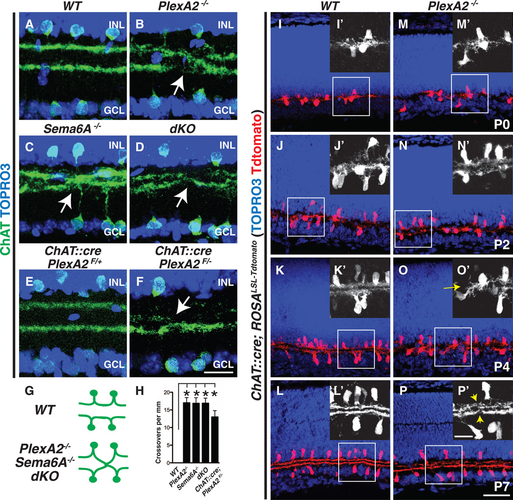
Fig. 2. Sema6A-PlexA2 signaling segregates On and Off SAC dendritic stratifications in vivo.
(A to D) Wild-type (A), PlexA2−/− (B), Sema6A−/− (C), and PlexA2−/−;Sema6A−/− (D) adult retina sections immunostained with antibodies to ChAT. On and Off SAC processes fail, with full penetrance, to completely segregate in PlexA2−/− (B), Sema6A−/− (C), and PlexA2−/−;Sema6A−/− (D) retinas (stratification crossovers indicated by white arrows, n ≥ 4 mice for each genotype). (E and F) Conditional removal of PlexA2 protein in SACs recapitulates PlexA2−/− stratification defects (n = 3 mice). (G) Schematics of SAC dendritic stratification in WT, PlexA2−/−, Sema6A−/−, and PlexA2−/−; Sema6A−/− mutants. (H) Quantification of SAC stratification crossovers in (A) to (D) and in (F) (n ≥ 6 retinas per genotype). Error bars, mean ± SD *P < 0.001. (I to P′) Characterization of SAC dendritic stratification in WT [(I) to (L′)] and PlexA2−/− [(M) to (P′)] retinas using the SAC Tdtomato genetic reporter. In WT retinas, On and Off SAC dendrites are intermingled at P0 [(I) and (I′)]. They then segregate during early postnatal retinal development [(J) and (J′)] and become completely separate stratifications by P4 [(K) and (K′)]. SAC dendritic stratifications in PlexA2−/− mutants [(M), (M′), (N), and (N′)] are indistinguishable from WT [(I), (I′), (J), and (J′)] at P0 and at P2. However, the stratification phenotype is observed by P4 in PlexA2−/− retinas [(O) and (O′), yellow arrow)] and persists through P7 [(P) and (P′), yellow arrowheads]. n = 3 mice for each genotype at each different developmental stage. Scale bars, 20 µm in (F) for (A) to (F), 50 µm in (P) for (I) to (P), and 20 µm in (P′) for (I′) to (P′).

